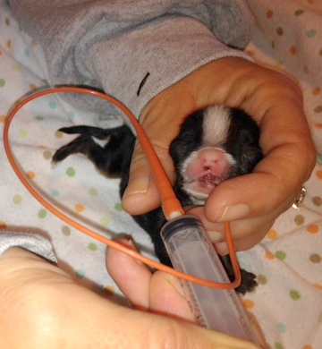
Caring for Cleft lip/palate pups
Homemade Puppy Formula
1 can evaporated milk
1/2-3/4 can distilled water (or baby water or boiled water-cooled)
1 cup plain yogurt (NOT fat free)
1 tbsp Karo syrup (corn syrup)
1 egg yolk
FEEDING:
Feed 1cc per OZ ( or 1 tbsp per pound) of body weight every 3 hrs - is the general rule of thumb.
Store in the refrigerator
Recipe using goats milk
10 ounces of goats milk
(I found it in my grocery store in the health food/Organic section)
1 tbsp. light corn syrup
1 cup of plain white (whole milk) NOT LOW FAT yogurt
1 egg yolk
Pour 10 oz. of goat's milk into a bowl and add in 1 tbsp. of corn syrup, 1 cup of plain white yogurt, and one egg yolk. Mix up the ingredients well using a whisk or electric mixer.
As a general rule, feed 1 cc. per 1 ounce body weight
If a puppy weighs 3 ounces, feed 3 cc's every three hours.
How to tube-feed a puppy
A puppy born with a cleft palate can't latch on properly to nurse! It may look like they are nursing but they may not be getting enough to sustain them. They may not be getting anything at all. Think of sucking on a straw with a hole in it. It looks like you are sucking but you are getting little to nothing.
A puppy born with a cleft palate should be pulled from mom immediately. If the puppy does manage to nurse just a little, they will be exposed to aspiration pneumonia. The safest way to feed a cleft palate baby is to tube-feed them. Below is a video showing how to tube-feed a puppy.
What you will need to care for a newborn pup with a cleft palate:
*A catheter feeding tube. They come in many sizes and can be purchased at your vets office
*Syringes
*A bulb syringe (to suck out babies mouth and nose if they burp up formula)
*A heating pad
*Hand-warmers for emergency trips to the vets office to keep baby warm
*A good scale (monitoring weight is essential!!)
*Puppy formula - either purchased at a pet store or homemade (recipes below)
*Nutri-cal or Karo syrup
Additional Information:
If your puppy shows signs of pneumonia (nasal discharge/lethargy/coughing,) bring them to the vet. Have a vaporizer on hand or a nebulizer.
Do NOT give a puppy weighing less than a pound antibiotics of ANY kind without consulting your veterinarian.
A compounded prescription will have to be made by a pharmacist! Full strength antibiotics on a newborn puppy will KILL the puppy.
It will shut down the liver and kidneys!!
The information provided is just a guide. I AM NOT A VETERINARIAN! I am only someone that has experience in tube-feeding.
Please see your vet for professional advice on how to care for any animal needing assistance.
Most vets will have no problem showing you how to tube-feed and if they do have a problem with it, find a new vet.
A cleft palate is a physical birth defect caused by incomplete fusing of the two halves of the palate during neonatal development. The palate is the roof of the mouth. The hard palate is the front part of the roof of the mouth, between the teeth, and separates the oral cavity from the nose. The soft palate extends from the back of the hard palate to the throat (pharynx) and is made of softer, fleshier tissue. When an animal swallows, the back of its soft palate normally swings up against the top of its throat, blocking the movement of air and food into the nasal cavity.
Some authorities classify cleft palates into two categories: 1) primary cleft palates, which involve the upper lip and/or the most frontal part of the hard palate; and 2) secondary cleft palates, which involve the middle to back parts of the hard and/or soft palates. A cleft lip is a congenital defect in which the two sides of the upper lip do not properly fuse in an unborn puppy. In the above classification scheme, a cleft lip is included within the definition of a primary cleft palate. Another name for this particular defect is harelip.
Causes of Cleft Palate
Cleft palates are caused by something that disturbs the normal processes that form the face and jaw during fetal development. There is a failure of fusion of certain paired structures in utero. The hard palate, soft palate or both can be affected. Most authorities think that there is a genetic cause; mating a male and a female that both have cleft palates reportedly results in more than 40% of their offspring also being affected with the condition. Poor maternal nutrition, viral infection of the bitch during pregnancy, exposure of the pregnant female to toxic drugs, chemicals or other substances, metabolic disorders and physical or mechanical interference with the fetus during development have all been implicated as potential causes of cleft palates in dogs. Administration of corticosteroids to the female during pregnancy has also been associated with cleft palates in her puppies.
Prevention of Cleft Palate
There appears to be a significant hereditary component to cleft palates in domestic dogs. One of the best ways to prevent this defect is to spay or neuter affected animals – or at least to remove them from the breeding population. Authorities recommend that corticosteroids or other drugs that can potentially cause birth defects not be given to bitches during pregnancy.
Symptoms of Cleft Palate in Dogs
Owners of dogs with cleft palates may notice one or more of the following things, especially in very young puppies:
Nasal congestion
Difficulty nursing or inability to suckle (caused by a physical inability to create enough suction or negative pressure to nurse properly)
Chronic nasal discharge (milk, mucus and/or pus dripping out of one or both nostrils of a newborn puppy)
Coughing
Gagging
Sneezing
Difficulty eating (dysphagia)
Difficulty breathing (dyspnea; respiratory distress; labored breathing; caused by aspiration/inhalation of milk, water or food)
Poor body condition; ill-thrift
Obvious “split upper lip”
Dogs at Increased Risk
Cleft palates occur sporadically in all breeds and mixed breeds, in dogs of either sex. However, they reportedly are more common in Beagles, Boston Terriers, Brittany Spaniels, Bulldogs (English and French), Cocker Spaniels, Dachshunds, German Shepherd Dogs, Labrador Retrievers, Pekingese, Pointers, Schnauzers, Shetland Sheepdogs and Shih Tzus. Brachycephalic breeds – those with broad skulls and short, flat faces - are predisposed to developing cleft palates.
How Cleft Palate is Diagnosed
Any newborn that is not thriving will be given a thorough physical examination by the attending veterinarian, who also will take a good history of the health of the bitch and of all of the littermates. A cursory examination of the upper lip and oral cavity by a veterinarian or knowledgeable breeder usually reveals a cleft palate, with the occasional exception being if the midline defect is isolated far back in the soft palate. This is not especially common in companion dogs. The normal initial database of blood work (a complete blood count and serum biochemistry profile) and a urinalysis may or may not be abnormal. If the puppy develops aspiration pneumonia as a result of a cleft palate, the preliminary blood work may identify an infection. Thoracic radiographs (chest X-rays) may be recommended if the puppy is showing signs of respiratory distress.
The best way to definitively diagnose a cleft palate is to conduct a thorough oral examination under general anesthesia. During that procedure, the veterinarian will be able to get a good look at the puppy’s hard and soft palates and evaluate the precise nature and extent of the defect.
Treatment Options
The goals of treating a cleft palate are to: 1) provide sufficient nutritional support until the puppy is old enough and healthy enough to survive surgery; 2) treat and resolve aspiration pneumonia or other associated respiratory disorders, if present; 3) close the anatomical defect surgically if possible; and 4) restore the dog’s ability to eat, drink and live normally, healthily and happily.
Intensive supportive care is usually necessary for any puppy with a cleft palate. The puppy’s physical inability to create enough suction to nurse normally usually requires that it be fed by hand. This involves carefully passing a flexible rubber tube down the dog’s esophagus (throat) directly into its stomach, and feeding either bitches’ milk or a milk-replacer formula through the tube, to ensure the puppy’s daily nutritional and caloric intake. This is not a particularly difficult procedure and most owners can be taught to tube feed their puppies at home. However, it is essential that the tube not be inserted down the trachea (wind pipe). Unfortunately, no matter how careful the person feeding the puppy is, puppies with cleft palates often develop aspiration pneumonia. Many of them simply do not thrive and die young.
Pneumonia must be treated and resolved before surgery is a realistic option. Antibiotic therapy may be necessary to manage secondary bacterial respiratory tract infections. Once the puppy is stable, surgical correction may be an option, especially if the palate defect is small. Reconstructive surgery is usually done at between 2 and 4 months of age, shortly after weaning. There are a number of different surgical techniques to rebuild a cleft palate, depending upon its location, but each of them is complex and should be performed only by a veterinarian experienced in the procedure. Multiple surgeries may be necessary to achieve successful correction, if that is even possible.
Caring for a newborn with a cleft palate is very time consuming and sometimes scary. If you don't think that you can provide adequate care, there is help available. Please contact us ASAP. We can mentor you or take the baby and provide care.

